How does mobile vet ultrasound work?
In the ever - evolving field of veterinary medicine, mobile vet ultrasound has emerged as a game - changer. As a leading supplier of Mobile Vet Ultrasound, I am excited to delve into how this technology works and its significance in animal healthcare.

The Basics of Ultrasound Technology
At its core, ultrasound technology relies on the principle of sound waves. Just like how bats use echolocation to navigate in the dark, ultrasound machines send out high - frequency sound waves into the body of an animal. These sound waves, which are above the range of human hearing (typically frequencies between 2 and 18 megahertz are used in veterinary ultrasound), travel through the tissues. When they encounter different types of tissues or structures within the body, such as organs, bones, or fluids, some of the sound waves are reflected back to the transducer, while others continue to penetrate deeper.
The transducer is a key component of the ultrasound machine. It is a handheld device that both emits the ultrasound waves and receives the reflected waves, known as echoes. The transducer contains piezoelectric crystals. When an electrical current is applied to these crystals, they vibrate and produce the ultrasound waves. Conversely, when the echoes return to the transducer, they cause the crystals to vibrate again, generating an electrical signal.
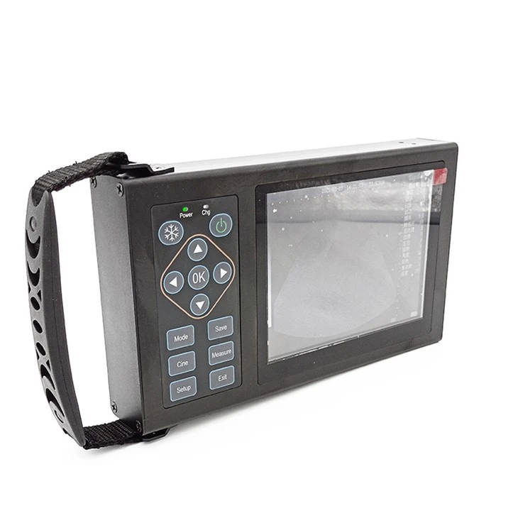
Mobile Vet Ultrasound Systems
Our mobile vet ultrasound systems are designed with portability and ease of use in mind. They are compact and lightweight, allowing veterinarians to bring the diagnostic power of ultrasound directly to the animal's location, whether it's on a farm, in a pet owner's home, or at a remote veterinary clinic.
These systems typically consist of a transducer, a control unit, and a display screen. The control unit processes the electrical signals received from the transducer. It uses complex algorithms to convert these signals into visual images that can be interpreted by the veterinarian. The display screen then shows these images in real - time, providing a live view of the internal structures of the animal's body.
How Mobile Vet Ultrasound is Used in Practice
Pregnancy Diagnosis
One of the most common applications of mobile vet ultrasound is pregnancy diagnosis in animals. For example, in livestock such as cows, mares, and sows, early and accurate pregnancy detection is crucial for proper herd management. With a Portable Ultrasound Scanner Veterinary Pregnancy, veterinarians can detect pregnancy as early as a few weeks after breeding.
During pregnancy, the developing fetus and associated structures, such as the placenta and amniotic fluid, have distinct ultrasound characteristics. The transducer is gently placed on the animal's abdomen, and the ultrasound waves penetrate the tissues to visualize the uterine contents. The presence of a gestational sac, embryo, or fetus can be clearly seen on the ultrasound image, allowing the veterinarian to confirm pregnancy and estimate the stage of gestation.
Musculoskeletal and Soft Tissue Evaluation
Mobile vet ultrasound is also valuable for evaluating musculoskeletal and soft tissue injuries in animals. In horses, for instance, it can be used to diagnose tendon and ligament injuries, which are common in athletic horses. The high - frequency ultrasound waves can provide detailed images of the tendons and ligaments, allowing veterinarians to detect tears, inflammation, or other abnormalities.
Similarly, in small animals like dogs and cats, ultrasound can be used to assess soft tissue masses, such as tumors or abscesses. By visualizing the size, shape, and location of these masses, veterinarians can make more informed decisions about further diagnostic tests and treatment options.
Cardiac Evaluation
Cardiac ultrasound, also known as echocardiography, is another important application of mobile vet ultrasound. In animals with heart problems, such as congenital heart defects or acquired heart diseases, echocardiography can provide valuable information about the structure and function of the heart.
The transducer is placed on the animal's chest in specific positions to obtain different views of the heart. The ultrasound waves allow the veterinarian to visualize the heart chambers, valves, and blood flow patterns. By measuring the size of the heart chambers, assessing the movement of the heart valves, and analyzing the blood flow velocities, veterinarians can diagnose heart diseases and monitor the progression of the condition over time.
Advantages of Mobile Vet Ultrasound
Real - Time Imaging
One of the major advantages of mobile vet ultrasound is its ability to provide real - time imaging. Unlike some other diagnostic imaging techniques, such as X - rays or CT scans, which provide static images, ultrasound allows veterinarians to observe the internal structures of the animal's body in motion. This is particularly useful for evaluating the function of organs, such as the heart and the digestive system.
Non - Invasive and Safe
Mobile vet ultrasound is a non - invasive and relatively safe diagnostic tool. It does not involve the use of ionizing radiation, which can be harmful to both the animal and the operator. This makes it a preferred choice for repeated examinations, especially in pregnant animals or young animals.
Cost - Effective and Accessible
Our mobile vet ultrasound systems are cost - effective compared to larger, stationary ultrasound machines. They also eliminate the need to transport animals to a specialized diagnostic facility, which can be time - consuming and stressful for the animals. This makes ultrasound more accessible to veterinarians in rural or underserved areas, as well as for those who provide mobile veterinary services.
Technical Considerations for Mobile Vet Ultrasound
Image Quality
While mobile vet ultrasound systems offer many advantages, ensuring high - quality images can be a challenge. Factors such as the type of transducer used, the depth of penetration, and the gain settings can all affect image quality.
The frequency of the transducer determines the resolution and penetration depth of the ultrasound waves. Higher - frequency transducers provide better resolution but have a shallower penetration depth, making them suitable for imaging superficial structures, such as the skin or the eyes. Lower - frequency transducers, on the other hand, can penetrate deeper into the body but have lower resolution. Therefore, choosing the appropriate transducer for the specific examination is crucial.
The gain settings on the ultrasound machine control the amplification of the echoes. Adjusting the gain correctly is important to ensure that the images are neither too bright nor too dark, allowing for accurate interpretation.
Training and Skill
Proper training and skill are essential for accurate interpretation of ultrasound images. Veterinarians need to have a good understanding of the normal anatomy and physiology of the animals they are examining, as well as the characteristic ultrasound appearances of different diseases and conditions.
Our company provides training and support to veterinarians who purchase our mobile vet ultrasound systems. This includes hands - on training sessions, online resources, and access to our technical support team. By investing in training, veterinarians can maximize the benefits of mobile vet ultrasound and provide better care for their patients.
Conclusion
Mobile vet ultrasound is a powerful diagnostic tool that has revolutionized the way veterinarians care for animals. Its portability, real - time imaging capabilities, and non - invasive nature make it an invaluable asset in veterinary practice. Whether it's for pregnancy diagnosis, musculoskeletal evaluation, or cardiac assessment, mobile vet ultrasound provides veterinarians with the information they need to make accurate diagnoses and develop appropriate treatment plans.
As a supplier of Mobile Vet Ultrasound systems, we are committed to providing high - quality products and excellent customer service. If you are a veterinarian interested in enhancing your diagnostic capabilities or a farm owner looking for more efficient animal healthcare solutions, we invite you to contact us to discuss your needs and explore our range of products. Our team of experts is ready to assist you in finding the right mobile vet ultrasound system for your practice.
References
- Bush, M., & Miller, R. E. (2018). Ultrasound in Small Animal Practice. Elsevier.
- Piermattei, D. L., & Flo, G. L. (2019). Surgical Anatomy of the Dog and Cat. Saunders.
- Thrall, D. E. (2017). Textbook of Veterinary Diagnostic Radiology. Elsevier.