What information can a cardiac veterinary ultrasound provide?
Cardiac veterinary ultrasound is a powerful diagnostic tool that provides invaluable information for veterinarians, allowing them to assess the structure and function of a animal's heart. As a veterinary ultrasound supplier, we understand the significance of this technology in veterinary medicine. In this blog, we'll explore the wealth of information that a cardiac veterinary ultrasound can offer.
Structural Information
One of the primary functions of cardiac veterinary ultrasound is to provide detailed images of the heart's structure. This includes the size, shape, and thickness of the heart walls. By examining these parameters, veterinarians can detect abnormalities such as hypertrophy (thickening of the heart muscle), dilation (enlargement of the heart chambers), or atrophy (wasting away of the heart muscle).
The ultrasound can clearly visualize the four chambers of the heart: the left and right atria and the left and right ventricles. Any irregularities in the size or shape of these chambers can indicate underlying heart conditions. For example, an enlarged left atrium may be a sign of mitral valve disease, while a dilated right ventricle could suggest pulmonary hypertension.
The heart valves are also clearly visible on a cardiac ultrasound. The mitral, tricuspid, aortic, and pulmonary valves can be examined for signs of damage, such as regurgitation (leaking) or stenosis (narrowing). Valvular heart disease is common in many animals, especially older dogs, and early detection through ultrasound can lead to more effective treatment and management.
In addition to the chambers and valves, the ultrasound can reveal the presence of any masses or tumors within the heart. These can be benign or malignant, and their early detection is crucial for determining the appropriate course of treatment. For example, a cardiac tumor may require surgical removal, chemotherapy, or radiation therapy, depending on its type and location.
Functional Information
Cardiac veterinary ultrasound not only provides information about the heart's structure but also its function. One of the key parameters that can be measured is the ejection fraction, which is the percentage of blood that is pumped out of the left ventricle with each heartbeat. A normal ejection fraction in dogs and cats is typically between 50% and 75%. A lower ejection fraction may indicate heart failure or other cardiac conditions.
The ultrasound can also assess the blood flow within the heart and the major blood vessels. Doppler ultrasound, a specialized technique, is used to measure the velocity and direction of blood flow. This can help detect abnormalities such as turbulent blood flow, which may be a sign of a heart murmur or a structural defect.
Another important aspect of cardiac function that can be evaluated is the contractility of the heart muscle. The ultrasound can show how well the heart is contracting and relaxing with each heartbeat. Reduced contractility may be a sign of cardiomyopathy, a disease that affects the heart muscle.
Assessment of Cardiac Rhythm
Cardiac veterinary ultrasound can also provide information about the heart's rhythm. By observing the movement of the heart chambers and the blood flow, veterinarians can detect arrhythmias, which are abnormal heart rhythms. Arrhythmias can be caused by a variety of factors, including heart disease, electrolyte imbalances, or certain medications.
Some arrhythmias may be benign and not require treatment, while others can be life - threatening. For example, ventricular tachycardia, a rapid and irregular heartbeat originating from the ventricles, can lead to cardiac arrest if not treated promptly. Early detection of arrhythmias through ultrasound can allow for timely intervention and better outcomes for the animal.
Detection of Congenital Heart Defects
Congenital heart defects are present at birth and can have a significant impact on an animal's health. Cardiac veterinary ultrasound is an essential tool for detecting these defects. Common congenital heart defects in animals include patent ductus arteriosus (PDA), ventricular septal defect (VSD), and atrial septal defect (ASD).
PDA is a condition where a blood vessel that should close shortly after birth remains open, allowing abnormal blood flow between the aorta and the pulmonary artery. VSD and ASD are holes in the walls separating the ventricles and atria, respectively. These defects can lead to abnormal blood flow patterns and put extra strain on the heart.

Early detection of congenital heart defects through ultrasound can help veterinarians determine the best course of treatment. In some cases, surgical repair may be possible, while in others, medical management may be the most appropriate option.
Monitoring of Treatment
Once a cardiac condition has been diagnosed, cardiac veterinary ultrasound is also useful for monitoring the effectiveness of treatment. For example, if a dog is being treated for heart failure with medications, regular ultrasound examinations can show whether the heart is responding to the treatment. The size of the heart chambers, the ejection fraction, and the blood flow patterns can all be monitored over time to assess the progress of the disease and the response to therapy.
If a surgical procedure has been performed to correct a congenital heart defect or a valvular problem, ultrasound can be used to ensure that the repair was successful and that there are no complications. This ongoing monitoring is crucial for providing the best possible care for animals with cardiac diseases.
Our Range of Veterinary Ultrasound Machines
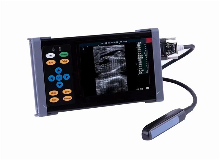
As a veterinary ultrasound supplier, we offer a wide range of high - quality ultrasound machines suitable for cardiac examinations. Our Portable Ultrasound Scanner Veterinary Pregnancy is a great option for on - the - go veterinarians or those working in small clinics. It is lightweight, easy to use, and provides clear images of the heart.
For equine practices, our Equine Ultrasound Machine is specifically designed to meet the unique needs of examining horses' hearts. It has a powerful transducer and advanced imaging capabilities to provide detailed and accurate information.
Our Laptop Ultrasound Machine combines the portability of a laptop with the functionality of a high - end ultrasound system. It is ideal for veterinarians who need a versatile and reliable machine for cardiac and other types of veterinary ultrasound examinations.
Conclusion
In conclusion, cardiac veterinary ultrasound is an indispensable tool in veterinary medicine. It provides a wealth of information about the structure, function, rhythm, and congenital defects of the heart. This information is crucial for accurate diagnosis, appropriate treatment, and ongoing monitoring of cardiac conditions in animals.
If you are a veterinarian looking to enhance your diagnostic capabilities or improve the care of your patients with cardiac diseases, we invite you to contact us to discuss our range of veterinary ultrasound machines. Our team of experts is ready to assist you in finding the right solution for your practice.
References
- Ettinger, S. J., & Feldman, E. C. (Eds.). (2010). Textbook of veterinary internal medicine: diseases of the dog and cat. Saunders Elsevier.
- Tilley, L. P., & Smith, F. W. K. (2012). Essentials of canine and feline electrocardiography. Wiley - Blackwell.
- Bonagura, J. D., & Twedt, D. C. (Eds.). (2018). Kirk's current veterinary therapy XV: small animal practice. Elsevier.